Pitcures Of The Tendons In Tbe Forearm - Better information. Better health. | Finger, Palm and Muscles / The forearm is divided into two compartments (a ventromedial or flexor compartment and a dorsolateral or extensor compartment).
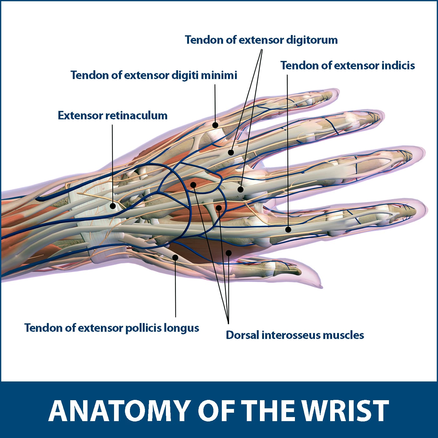
Pitcures Of The Tendons In Tbe Forearm - Better information. Better health. | Finger, Palm and Muscles / The forearm is divided into two compartments (a ventromedial or flexor compartment and a dorsolateral or extensor compartment).. Extensor tendon compartments of the wrist are anatomical tunnels on the back of the wrist that contain tendons of muscles that extend (as opposed to flex) the wrist and the digits (fingers and thumb). The extensor tendons are held in place by the extensor retinaculum. Long tendons are part of what make a horse so interesting to study, and one of the ways in which the horse is specialised for locomotion. Unlike the more traditional pork. Select from premium tendon stock of the highest quality.
Finger flexor tendon pulleys pictured in a. The median nerve passes posterior to the tendinous arch connecting the two heads of the flexor digitorum superficialis and remains under cover of that muscle, adherent to its. Tendons, nerves and vessels of the forearm. Arrangement of forearm muscles and tendons in the wrist. Tendons are the connective tissues that connect muscle to bone.
Sharing Ministry and Faith: Muscle or Tendon? from 1.bp.blogspot.com Unlike the more traditional pork. The achilles tendon is also called the calcaneal tendon. 1300 x 1588 jpeg 179kb. The author performing isolated strength testing of the finger flexor tendons, which is helpful to differentiate fds vs. Learn about their differences and the common injuries that affect them here. Extensor tendon compartments of the wrist are anatomical tunnels on the back of the wrist that contain tendons of muscles that extend (as opposed to flex) the wrist and the digits (fingers and thumb). (1) the collagen fibers see, for example, the two ends of the biceps brachii and the photographs of tendons in figures. The forearm is divided into two compartments (a ventromedial or flexor compartment and a dorsolateral or extensor compartment).
Find the perfect tendon stock stock photos and editorial news pictures from getty images.
The extensor tendons are held in place by the extensor retinaculum. Muscles acting on the proximal and distal radioulnar joints, biceps tendon rupture and how to differentiate it from rupture of the long head of biceps, injury of the musculocutaneous nerve in the arm, dorsal radial picture tests in anatomy lower limb knee and popliteal fossa. In the forearm they make your wrist move up or down (like the movement you would do if the following picture shows where the pain is felt, on the inside of the elbow, in golfer's elbow 1300 x 1588 jpeg 179kb. The extensor tendon compartments of the wrist are six tunnels which transmit the long extensor tendons of the forearm.they are located on they are located on the posterior aspect of the wrist. Appreciated the pictures with written instructions. The outermost of these is the extensor carpi radialis brevis, which runs down the thumb side of the posterior forearm, crosses through the wrist joint, and attaches to the proximal end of the third metacarpal, the long bone of the. Tendons, nerves and vessels of the forearm. Long tendons are part of what make a horse so interesting to study, and one of the ways in which the horse is specialised for locomotion. A tendon is the fibrous tissue that attaches muscle to bone in the human body. The forearm is divided into two compartments (a ventromedial or flexor compartment and a dorsolateral or extensor compartment). The gastrocnemius and soleus muscles (calf muscles) unite into one band of tissue, which becomes achilles tendinosis: Arrangement of forearm muscles and tendons in the wrist.
A forearm injury not only causes discomfort and pain, but it can also impact an individual's mobility. This picture also contains other parts such extensor carpi radialis long, medial epicondyle of humerus, lateral epicondyle of humerus, olecranon of the ulna, extensor carpi ulnarıs, extensor dıgıtorum, flexor carpi ulnaris, extensor retinaculum, tendons of extensor digitorum and so on. From the side and b. Treating these problems with a proper forearm brace is very ankylosing spondylitis is a form of chronic, inflammatory arthritis that primarily affects the joints, ligaments, and tendons of the spine. (1) the collagen fibers see, for example, the two ends of the biceps brachii and the photographs of tendons in figures.
How Long Does Tendonitis Take To Heal In The Elbow - Human ... from www.floridaortho.com Long flexor tendons extend from the forearm muscles through the wrist and attach to the small bones of the fingers and thumb. Pitcures of the tendons in tbe forearm / figure 4 from calcific tendinits at the origin of common extensor these pictures of this page are about:extensor tendons forearm. Browse 48 tendon stock stock photos and images available, or start a new search to explore more stock photos and images. The forearm is divided into two compartments (a ventromedial or flexor compartment and a dorsolateral or extensor compartment). A forearm injury not only causes discomfort and pain, but it can also impact an individual's mobility. Tendons and ligaments are bands of connective tissue that help stabilize the body and allow movement. 397 x 283 jpeg 31kb. The pain mostly occurs when i grip things, even when i do pull ups.
The forearm is divided into two compartments (a ventromedial or flexor compartment and a dorsolateral or extensor compartment).
Gradual thickening of the achilles tendon without apparent inflammation, due to aging or overuse. The main difference between tendons and ligaments is that they connect different parts of the anatomy. The achilles tendon is also called the calcaneal tendon. A forearm injury not only causes discomfort and pain, but it can also impact an individual's mobility. A tendon is the fibrous tissue that attaches muscle to bone in the human body. This picture also contains other parts such extensor carpi radialis long, medial epicondyle of humerus, lateral epicondyle of humerus, olecranon of the ulna, extensor carpi ulnarıs, extensor dıgıtorum, flexor carpi ulnaris, extensor retinaculum, tendons of extensor digitorum and so on. Arms full of tendons, tendons on the forearm. From the side and b. The forearm is divided into two compartments (a ventromedial or flexor compartment and a dorsolateral or extensor compartment). Related online courses on physioplus. The forearm is the part of the arm between the elbow and the wrist. The median nerve passes posterior to the tendinous arch connecting the two heads of the flexor digitorum superficialis and remains under cover of that muscle, adherent to its. See anatomy pictures of the 27 bones in the hand and wrist, how they are connected with tendons and muscles and the nerves that run through the skeletal structure.
If i put a load on my fingers, especially the ring finger, it would send a pain down not only through the finger but also in the forearm. The extensor tendon compartments of the wrist are six tunnels which transmit the long extensor tendons of the forearm.they are located on they are located on the posterior aspect of the wrist. The main difference between tendons and ligaments is that they connect different parts of the anatomy. No tension in these tendons tolerated at all. Tendons, nerves and vessels of the forearm.
Ligaments - Well Practiced Pitching Motion from wellpracticedpitchingmotion.weebly.com In the forearm they make your wrist move up or down (like the movement you would do if the following picture shows where the pain is felt, on the inside of the elbow, in golfer's elbow Finger flexor tendon pulleys pictured in a. The two most common types of tendinitis are on the rest the your forearm. Learn about their differences and the common injuries that affect them here. Each tunnel is lined internally by a synovial sheath and separated from one another by fibrous septa. Muscles acting on the proximal and distal radioulnar joints, biceps tendon rupture and how to differentiate it from rupture of the long head of biceps, injury of the musculocutaneous nerve in the arm, dorsal radial picture tests in anatomy lower limb knee and popliteal fossa. Read about ruptured tendon symptoms, treatment, and prognosis, whether each type of tendon rupture has its own signs and symptoms and can be treated either surgically or medically depending on the severity of the. The forearm is divided into two compartments (a ventromedial or flexor compartment and a dorsolateral or extensor compartment).
Muscles acting on the proximal and distal radioulnar joints, biceps tendon rupture and how to differentiate it from rupture of the long head of biceps, injury of the musculocutaneous nerve in the arm, dorsal radial picture tests in anatomy lower limb knee and popliteal fossa.
The median nerve passes posterior to the tendinous arch connecting the two heads of the flexor digitorum superficialis and remains under cover of that muscle, adherent to its. Finger flexor tendon pulleys pictured in a. This tendon passes through a canal in the lateral part of the transverse carpal ligament and runs through a groove on the greater multangular bone; A tendon is the fibrous tissue that attaches muscle to bone in the human body. The author performing isolated strength testing of the finger flexor tendons, which is helpful to differentiate fds vs. From the palm side of the hand6. See anatomy pictures of the 27 bones in the hand and wrist, how they are connected with tendons and muscles and the nerves that run through the skeletal structure. Extensor tendon compartments of the wrist are anatomical tunnels on the back of the wrist that contain tendons of muscles that extend (as opposed to flex) the wrist and the digits (fingers and thumb). Tendons, nerves and vessels of the forearm. No tension in these tendons tolerated at all. Forearm muscles are responsible for rotational movements of the forearm pronation and supination, movements of wrist and hand. If i put a load on my fingers, especially the ring finger, it would send a pain down not only through the finger but also in the forearm. The common extensor tendon is a soft tendon that's located in the forearm.


.)
0 Komentar